NIS3D: A Completely Annotated Benchmark for Dense 3D Nuclei Image Segmentation
Abstract
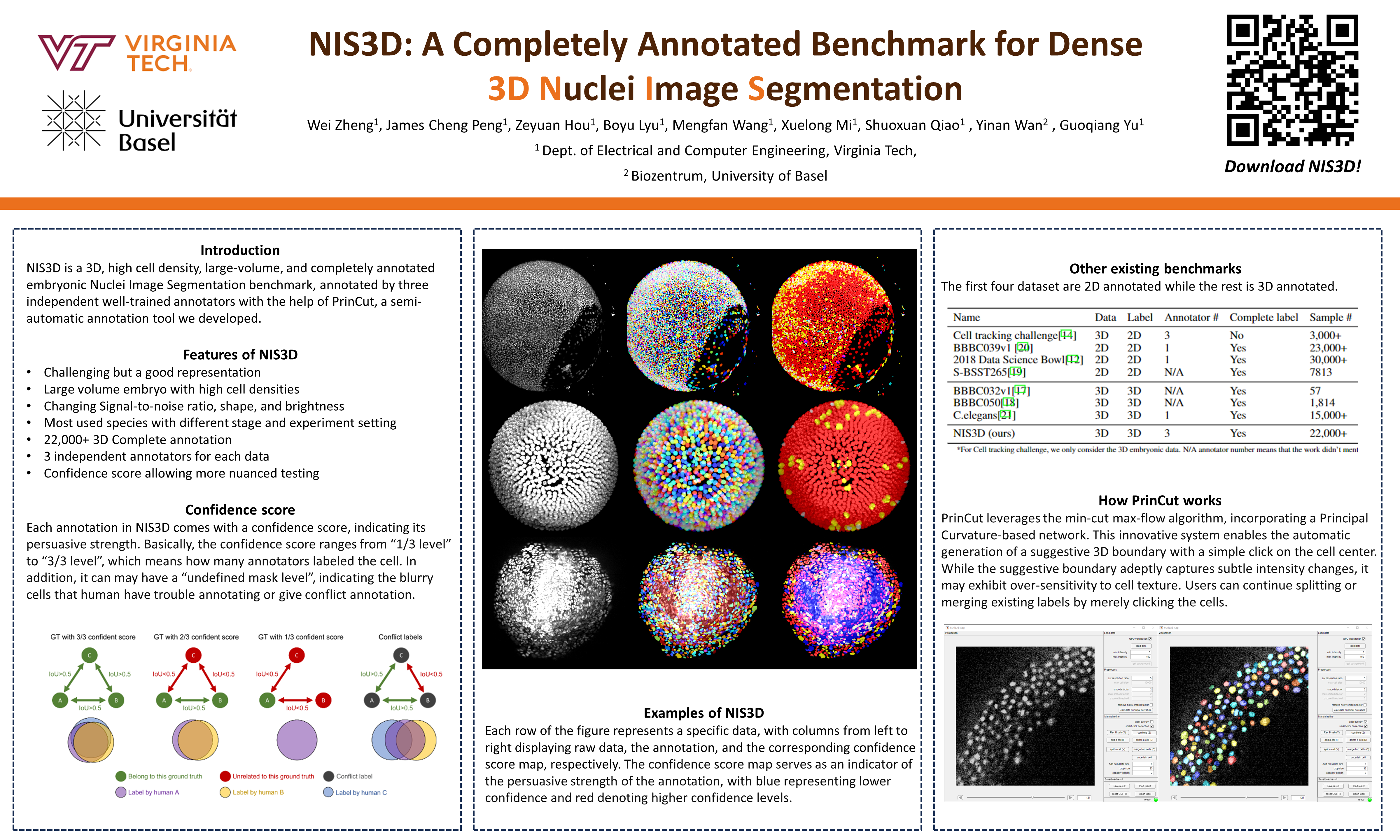
3D segmentation of nuclei images is a fundamental task for many biological studies. Despite the rapid advances of large-volume 3D imaging acquisition methods and the emergence of sophisticated algorithms to segment the nuclei in recent years, a benchmark with all cells completely annotated is still missing, making it hard to accurately assess and further improve the performance of the algorithms. The existing nuclei segmentation benchmarks either worked on 2D only or annotated a small number of 3D cells, perhaps due to the high cost of 3D annotation for large-scale data. To fulfill the critical need, we constructed NIS3D, a 3D, high cell density, large-volume, and completely annotated Nuclei Image Segmentation benchmark, assisted by our newly designed semi-automatic annotation software. NIS3D provides more than 22,000 cells across multiple most-used species in this area. Each cell is labeled by three independent annotators, so we can measure the variability of each annotation. A confidence score is computed for each cell, allowing more nuanced testing and performance comparison. A comprehensive review on the methods of segmenting 3D dense nuclei was conducted. The benchmark was used to evaluate the performance of several selected state-of-the-art segmentation algorithms. The best of current methods is still far away from human-level accuracy, corroborating the necessity of generating such a benchmark. The testing results also demonstrated the strength and weakness of each method and pointed out the directions of further methodological development. The dataset can be downloaded here: https://github.com/yu-lab-vt/NIS3D.